


September 5, 2024
5 Minimum read time
Scientists have made the skin of living mice transparent using a simple edible dye.
A new study used the superabsorbent dye tartrazine, commonly used as the common food coloring Yellow No. 5, to make tissues in living mice transparent, temporarily revealing the animals’ internal organs and blood vessels.
Skin normally scatters light, a phenomenon represented by the white lines at the beginning of this video. However, when Yellow No. 5, a food, drug, and cosmetic dye, is absorbed into the skin, scattering is reduced and light penetrates deeper, making the tissue transparent. (This technology has not been tested on humans. Dyes can be harmful. Always use caution when using dyes and do not ingest them directly, apply them to people or animals, or misuse them in any other way.)
Keyi “Onyx” Li/National Science Foundation
In just a few minutes, applying a common food dye to a mouse can make any part of its skin as transparent as glass.
In a study published today, science, Researchers sprayed live mice with a solution of the dye tartrazine, a common coloring agent in foods, drugs, and cosmetics. Clean the organization— creating temporary windows that reveal the organs, muscles, and blood vessels of the body. The procedure is a new form of technology known as “optical tissue clearing,” and while it has not yet been tested on humans, it could one day provide a way to view and monitor injuries or diseases without special imaging equipment or invasive surgery.
“One of the unique things about our strategy is that we directly change the optical properties of the tissue,” says Jihao Ou, a physicist at the University of Texas at Dallas and the study’s lead author.
About supporting science journalism
If you enjoyed this article, please support our award-winning journalism. Subscriptions. By purchasing a subscription, you help ensure a future of influential stories about the discoveries and ideas that shape our world today.
Skin, like most mammalian tissues, is highly opaque because its mixture of water and densely packed lipids, proteins, and other essential molecules scatters light in all directions. “The concept is similar to bubbles of water,” Ou explains. “If you have water and air, they’re both transparent on their own. But when you mix the two, you end up with microscopic bubbles that are no longer transparent.” Think of a rushing stream or a crashing wave. The variation in transparency is caused by the different refractive indices—the amount of light that bends as it passes through an object or substance—of water and air molecules. The fats and proteins in rodent and human skin typically have higher refractive indices than water, creating a contrast that’s hard to see. In the new study, Ou and his colleagues looked for light-absorbing molecules that could make the various refractive indices within the skin layer more similar, essentially reducing the amount of light scattered across the entire surface.

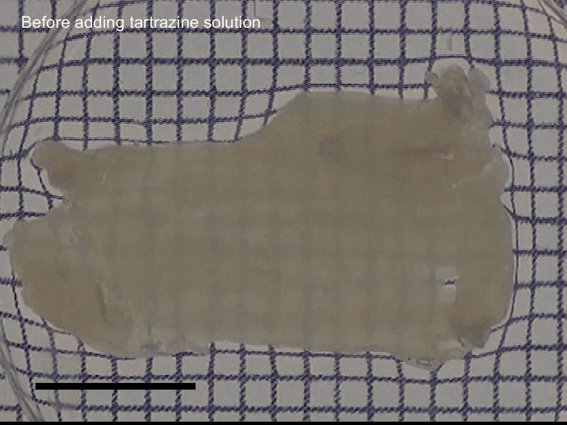
Photograph of a “scattering phantom”, a sample that mimics the optical distribution of human tissue made of agarose hydrogel. The phantom contains increasing concentrations of tartaric acid and the same concentration of silica particles. The scale bar represents 5 millimeters.
Guosong Hong/Stanford University
The team tested 21 different synthetic dyes before finding the highly absorbent one, tartrazine, commonly known as Yellow No. 5. The refreshing lemon yellow dye is approved by the Food and Drug Administration for limited use in foods, drugs, and cosmetics. It’s commonly found in chips, sodas, candy, butter, vitamins, and pills. Tartrazine makes the refractive index of the molecules it encounters more uniform, allowing red and yellow light to pass through, similar to the colors of the underlying tissues. At the same time, the dye absorbs most of the light in the near-UV and blue spectrum wavelengths, reducing the scattering of this type of light. “The higher the absorption, the more efficient the molecule,” Ou explains. FDA restrictions on chemicals and additives in food have led the food industry to look for “highly efficient chemicals,” even in small amounts.
Animated stills from real-time imaging video show the dynamic process of chicken breast tissue changing from opaque to transparent after immersion in a 0.6 molar solution of tartazan (yellow No. 5). The progression is shown before application of the tartazan solution and ranges from 0.2 to 60 seconds after. The scale bar represents 5 mm.
Guosong Hong/Stanford University
The researchers tested different concentrations of the dye on a “dispersion phantom”—a square sample that mimics the optical distribution of human tissue—and on pieces of raw chicken breast. They then gently massaged the dye onto the skin of anesthetized rats, where it was absorbed like a “facial cream.” In less than 10 minutes, the team began to see internal features beneath the top layer of tissue under visible light. Rubbing tartarzine on the animals’ stomachs produced activity in the digestive tract, and applying it to exposed muscle on one of their legs revealed the digestive tract. Using high-resolution laser imaging, the scientists were able to see nerves in the stomach, small units of muscle called sarcomeres, and even the vascular structure of the brain when the dye was applied to the rats’ scalps. If the tartarzine was not washed off, its effects lasted for about 10 to 20 minutes before the skin returned to its original state.

Still images from real-time video demonstrate the optical transparency of the mouse abdomen, allowing visualization of the animal’s abdominal organs. Scale bar represents 5 mm.
“Achieving optical transparency in living animals using absorbing molecules”, Zihao Ou et al. scienceVol. 385. Published online September 5, 2024
Past research They focused on introducing already transparent substances, including glycerol and fructose solutions that made the skin transparent. These molecules could also reduce light scattering, but “they weren’t very effective. [as tartrazine] “That’s because they’re not ‘colored’ enough,” says Guosong Hong, a materials science engineer at Stanford University and senior author of the new paper. Other approaches that remove essential molecules from tissues instead of adding new ones can achieve similar effects, but they can only be done in nonliving animals or biopsied tissues. For example, Rajan Kulkarni, a dermatologist at Oregon Health & Science University, led the Optical Tissue Clearing Project in 2014. Completely dissolved the entire organs and lipids of the animal. And we replaced it with a transparent hydrogel. “That was always the limitation, and something was needed. In vitro“We had to remove tissue or remove organs, otherwise the organism itself was no longer alive,” says Kulkarni, who was not involved in the new study. “This method [in the new paper] This is interesting because it can pass through the skin or epidermis. [in living animals] “Make it transparent so I can visualize what’s underneath.”
Although it’s still a long way from human testing, the concept could one day have useful medical applications. Dr. Hong suggests that it could potentially aid in the early detection of skin cancer and make laser-based tattoo removal simpler. It could also make veins more visible for drawing blood or administering fluids with a needle, especially in elderly patients whose veins are difficult to find. In some cases, this strategy could be a more attractive option than using imaging techniques such as magnetic resonance imaging (MRI) and ultrasound. “I can definitely see how this could be useful for visualization experiments in mice and other types of animals, because it gives you the ability to visualize at optical microscopy resolution, whereas other MRI, CT, [computed tomography]“Ultrasound doesn’t resolve that fine,” Kulkarni said. “From a proof-of-concept standpoint, it’s fantastic. Clinically, we don’t know yet.”
The researchers did not observe any adverse effects in mice after removing the dye. However, Ou says more effective molecules similar to tartadine need to undergo further testing for human safety. Tartadine can cause: allergic reaction. And while the coloring is FDA-approved, the agency has strict restrictions. Amount used in product. In this study, the mice could tolerate the highest concentration used, 0.6 molar, for a short test period. However, “human skin is about 10 times thicker than that of humans. [that of] “In mice, that means the time it takes to diffuse is much longer. What might be a few minutes in a mouse could be hundreds of minutes in a human,” Ou says. “We hope that this initial work will lead to more follow-up efforts suggesting new molecules that are more effective and safer for humans.”